(last updated on 7th June 2026)
If you would like to support this freely accessible database, you can do so by purchasing the book →
Although each micrometeorite is unique in its overall appearance, micrometeorites can still be classified or grouped on the basis of characteristic features.
In the database https://www.micrometeorites.org/database, such information from surface analyzes of several hundred micrometeorites (mainly cosmic spherules) as well as light photos, scanning electron images and EDX data are available. For some micrometeorites there is also information from sectioned and polished parts thus giving insights into the interior of the particle. The selection of the filter criteria does not follow any recognized guidelines but my personal selection.
About 100 terrestrial particles (clearly marked as such) were added to allow a comparison between micrometeorites and terrestrials.
The data can be filtered in the selection view according to selected characteristics. In the single view (detailed view), all available information is compiled for each micrometeorite.
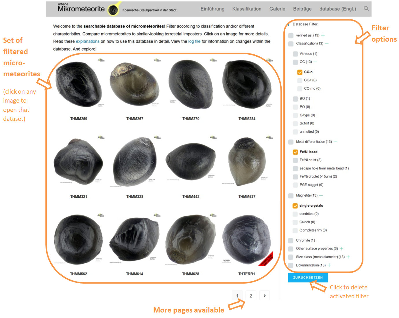
Selection View
The selection view gives an impression of the appearance and diversity of the micrometeorites, depending on the selected filter. In addition it allows to compare cosmic and terrestrial particles. The view offers the following options (see image below):
- Click on an image or on the ID of a micrometeorite in the displayed list to open it in the single view and get detailed information
- Switch the page if there are more micrometeorites in the selection than show on the screen
- Use the filter to define your selection (explanation on the filter options see below)
- Reset filters
Single View
The single view gives insight into the details of the selected particle. The following options then open up (see image below):
- Activate image series by clicking on the magnifying glass
- Hover the cursor over the active image to magnify it
- Click on any small image to make it the active image
- Click on the arrows in the top right corner to go to the previous or subsequent dataset

Using database filter
By using the filter you can modify the selection of micrometeorites displayed in the selection view. The particles criteria are:
- Verified as:
- Classification
- Metal differentiation
- Magnetite appearance
- Chromite crystals
- Various other surface properties
- Size class
- Available documentation
Their sub-categories (e.g. „Fe / Ni bead“) are primarily used as selection criteria. The superordinate categories „Classification“, „Metal differentiation“ etc. are only used for grouping these selectable criteria. Clicking on the plus sign to the right of the category name opens its sub-categories, clicking on a minus sign closes them again.
Several selection criteria can be combined. An „and“ link applies here, i. H. the selection is limited to micrometeorites that meet all of the selected criteria.
The number of available data records (depending on the current selection) is displayed in brackets behind each selection criterion.
The selection criteria are explained in detail and examples are given below.
Verified as:
With this filter, the selection can be limited to micrometeorites or terrestrial particles, respectively. Without setting this filter, both are displayed and can thus be compared with each other. Micrometeorites are listed first.
Since terrestrial particles are provided in the database with the different properties of micrometeorites according to their similarity, their selection also changes when using the different filter criteria.
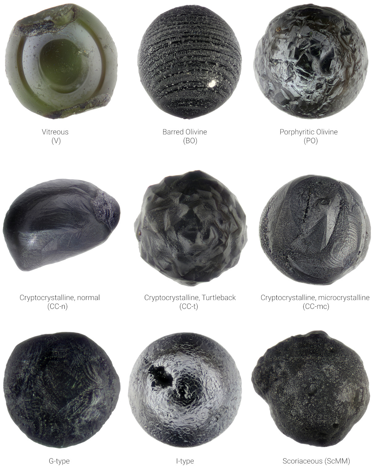
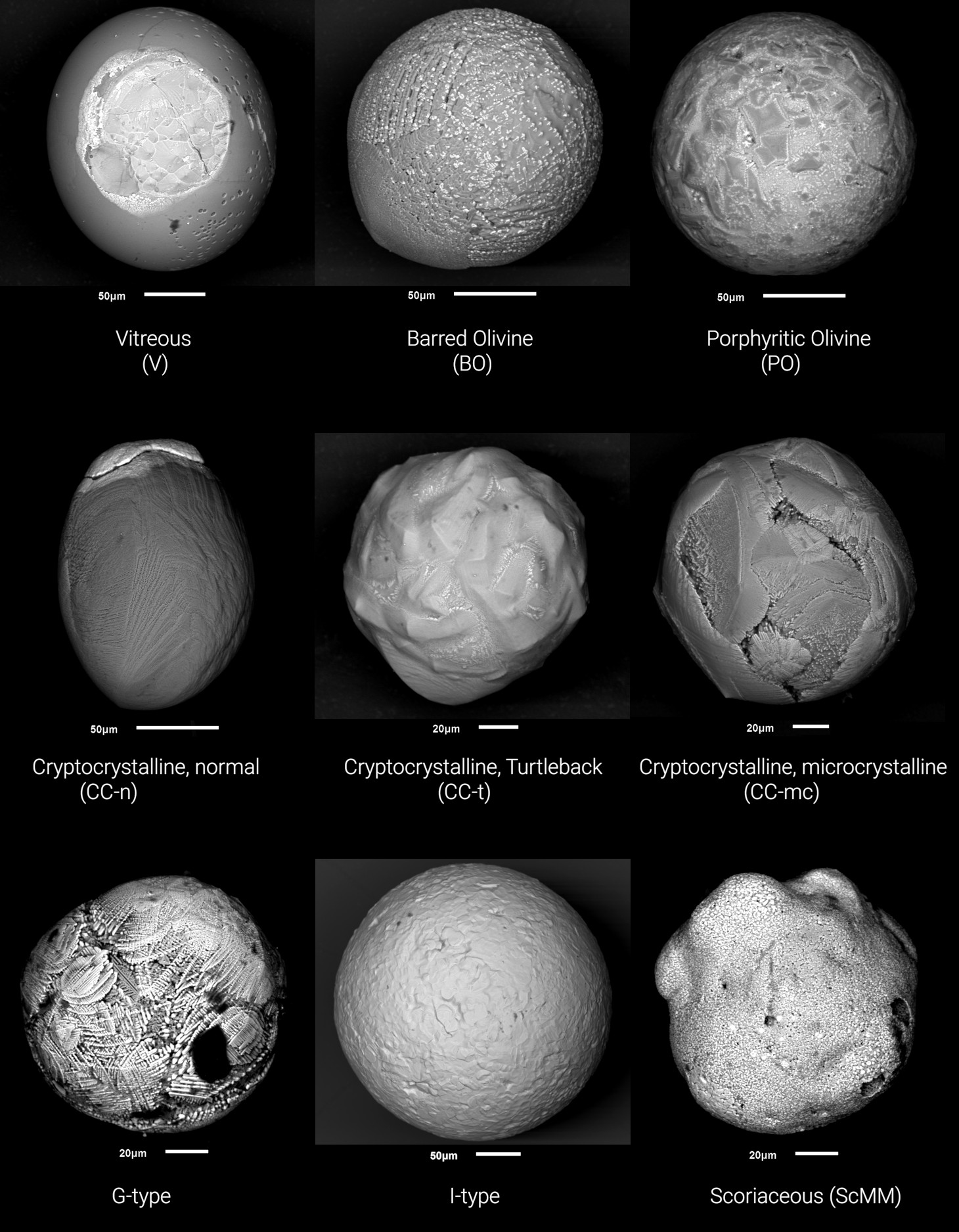
Classification
The following figure shows a typical representative of each of the classification sub-categories that occur. The classification is based on Genge et al. 2008 and regarding the subdivision of type CC according to Suttle and Folco 2020 (see also here). It should be noted, however, that the variation is quite high and there are particles that are difficult to assign in the classification based on surface characteristics only.
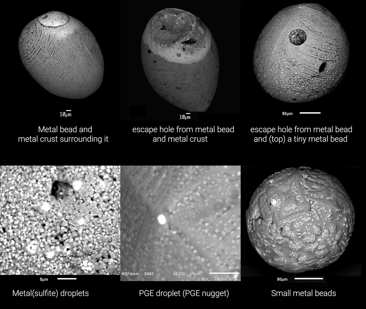
Metal differentiation
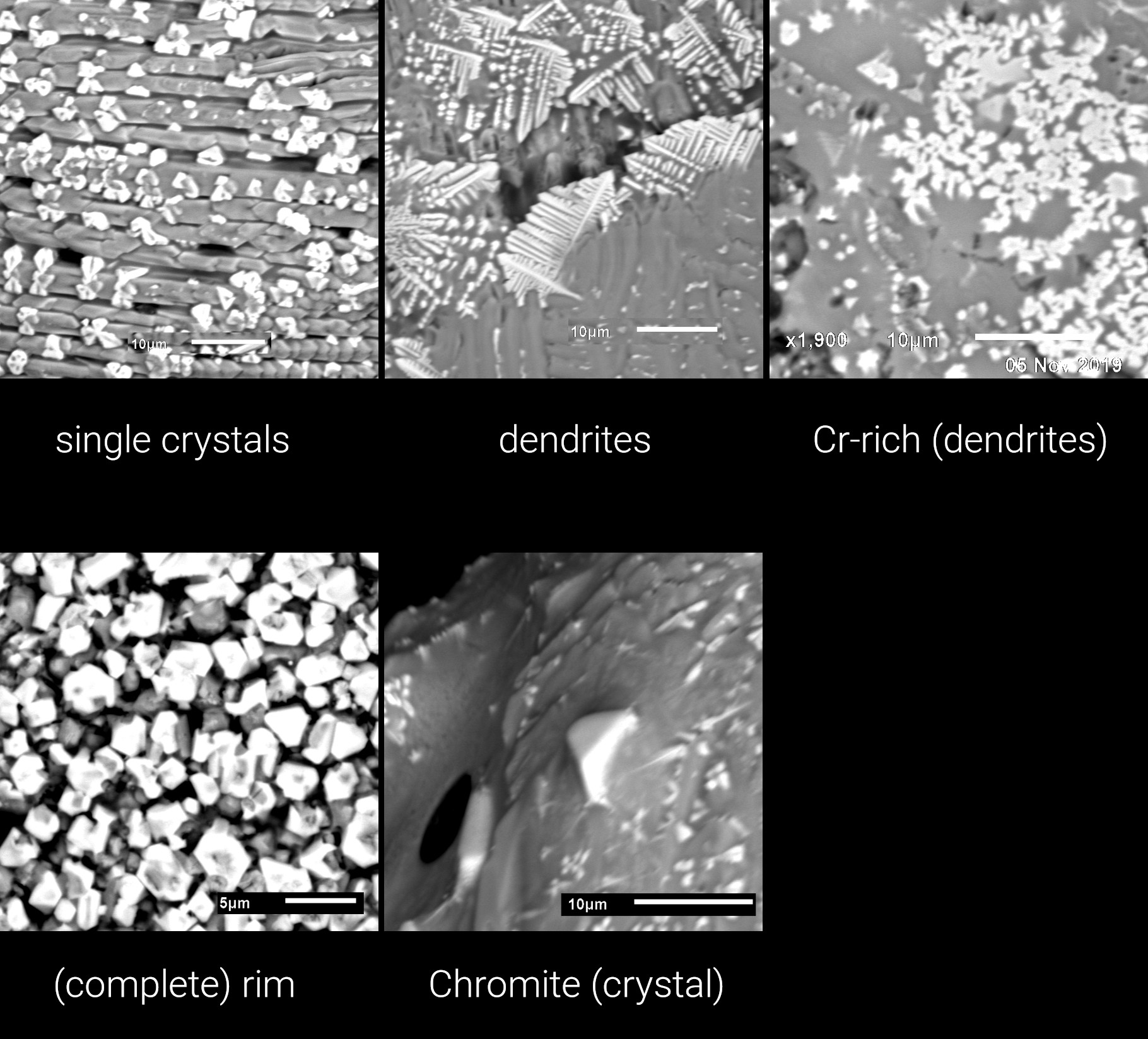
When the particle melts as it passes through the Earth’s atmosphere, the metal and silicate phases often separate. After solidification, these appear as metal spheres or metal droplets on the outer edge of the silicate body or as exit holes when the metal droplet has completely detached from the particle. Metal beads contain mainly iron (Fe) and often also nickel (Ni), in smaller amounts sometimes cobalt (Co) or other small admixtures of elements. The degree of oxidation varies. The following graphic with SEM-BSE images shows some representatives.
Appearance of magnetite and chromite
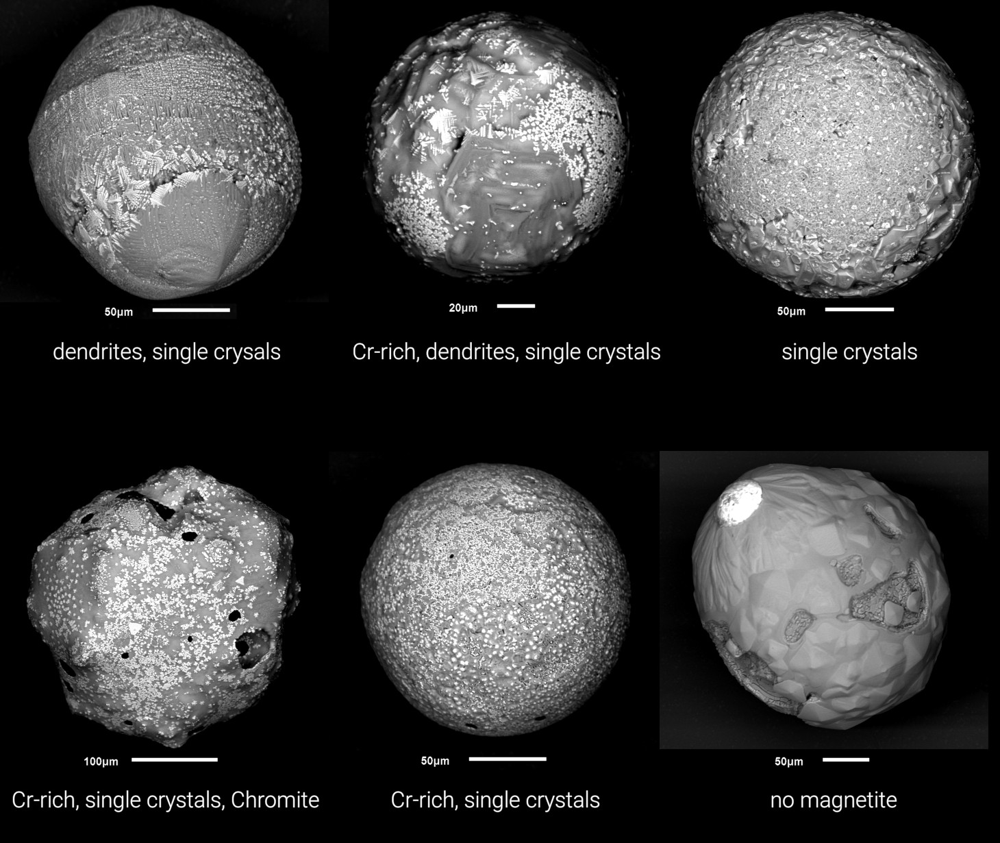
Magnetite almost always forms at the surface of cosmic spherules and other micrometeorites when iron gets in contact with atmospheric oxygen and it oxidites. Depending on the speed of this process and the chromium content of the particle, different shapes can result, which are shown in the following graphic. Chromite crystals form at particularly high Cr contents, but could also be relics of the unmelted material.
Surface properties
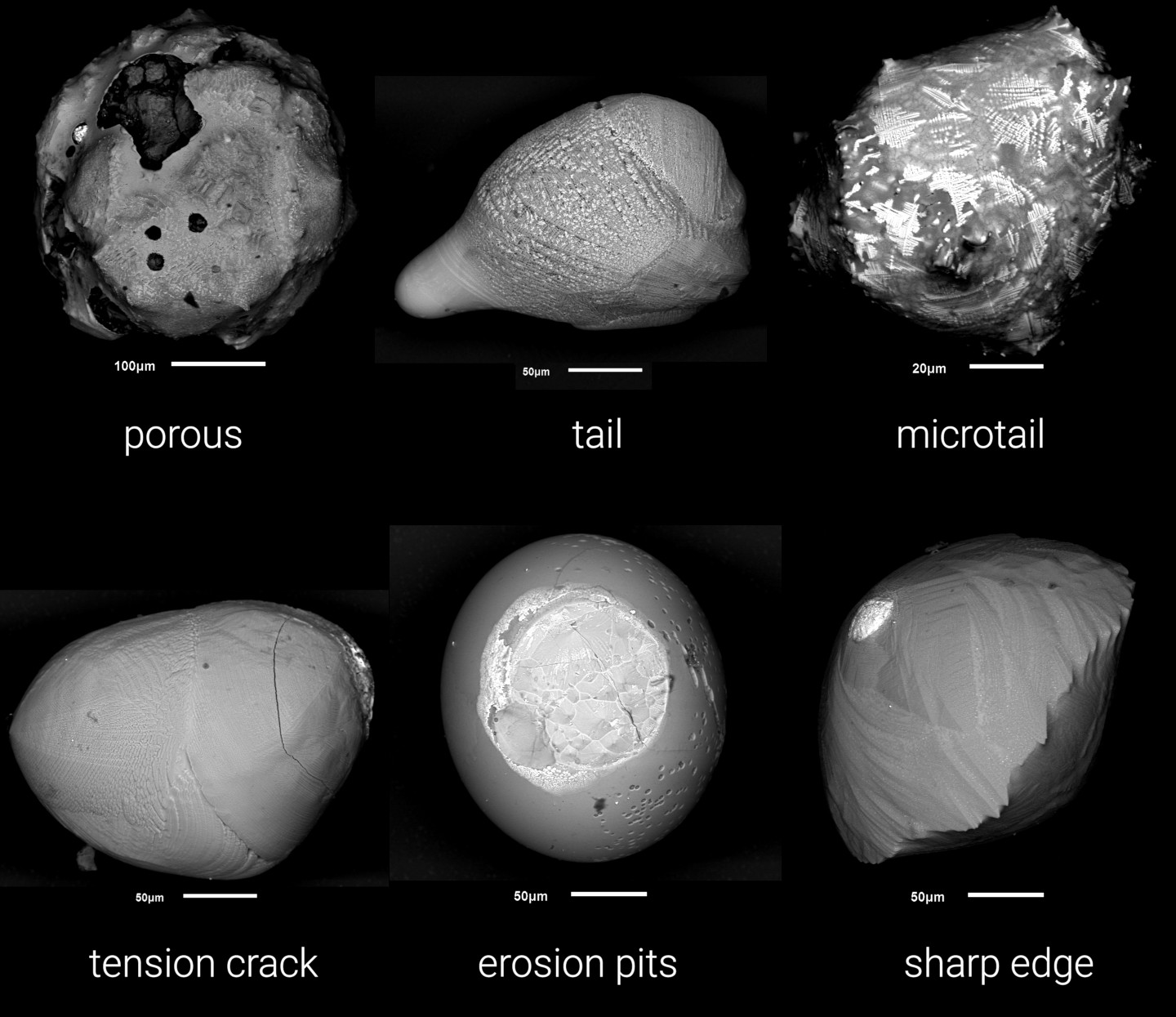
Here a choice of various other frequently observed characteristics are summed up (see figure below).
Size class mean
These categories divide the micrometeorites into certain size classes, according to which they can be filtered.
Documentation
This defines which information about the particle is available in the database. Mostly these are:
- light image: using a microscope, digital camera, ring lighting and photo stacking
- SEM image: backscattered electron (BSE) imaging at scanning electron microscope (SEM)
- EDX data: chemical analysis by energy dispersive X-ray spectrometry (EDX, EDS)
- sectioned: SEM image: backscattered electron (BSE) imaging at scanning electron microscope (SEM) of sectioned and polished particles set in epoxy resin
- sectioned: EDX data: chemical analysis by energy dispersive X-ray spectrometry of polished particle interiors (EDX, EDS)
Now feel free to explore the database!